 Medorax
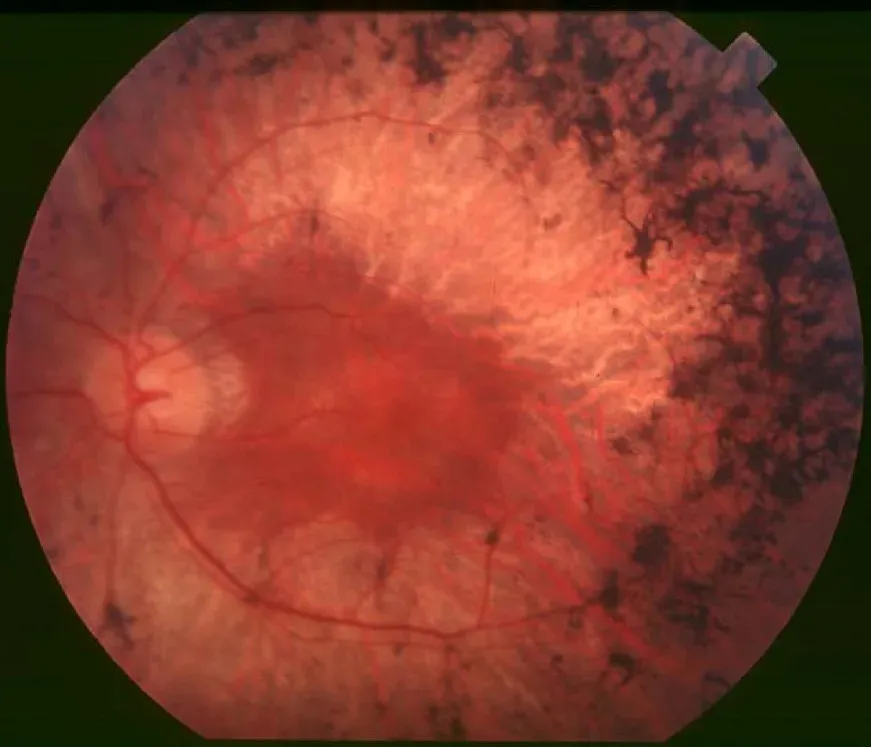
Retinosis pigmentaria
Fondo de ojo · Mácula y retina
Hallazgos
Espículas óseas de pigmento en la periferia, arteriolas adelgazadas y disco pálido céreo.
Interpretación
Retinosis pigmentaria: distrofia hereditaria de fotorreceptores (bastones primero).
Perla clínica
Clínica: ceguera nocturna y pérdida progresiva del campo periférico (visión en túnel). Puede asociarse a síndromes (Usher, Bardet-Biedl).
Medorax es material educativo y de repaso. No es un producto sanitario, no diagnostica ni indica tratamientos, y no sustituye la formación médica reglada, las guías clínicas vigentes ni el criterio de un profesional de la salud.
- Categoría
- Mácula y retina
- Dificultad
- Avanzado
- Tipo
- Retinografía
Origen: Christian Hamel · CC BY 2.0 · Wikimedia Commons · Licencia CC BY 2.0
Descargar Medorax